The key difference between apical and radial pulse is that apical pulse is a pulse site situated on the left side of the chest, over the apex of the heart, while radial pulse is one of the peripheral pulse sites located on the lateral of the wrist.
Jan 13, 2020 The apical pulse refers to the pulsation at the apex of the heart. The heart in a healthy person is situated such that the apex is in the left part of the chest, pointing down and to the left. It is also sometimes referred to as the “point of maximum impulse”, or PMI. About Press Copyright Contact us Creators Advertise Developers Terms Privacy Policy & Safety How YouTube works Test new features Press Copyright Contact us Creators. The apical pulse is the point of maximal impulse and is located at the apex of the heart. If you look at the heart in the body, it is flipped upside down with the base being at the top and the apex at the bottom. Video Demonstration on How to Assess the Apical Pulse How to Assess the Apical Pulse.
The heart is the organ that pumps blood across the body. Moreover, it purifies the blood and delivers nutrients to our body tissues and organs. Heart functioning can be detected by the sounds of the heart valve opening and closing. Abnormal heart rate indicates the possibility of heart diseases, heart failure or an overactive thyroid gland. Pulse is the heart rate – the vibration of blood as your heart pumps. It can be measured by the apical pulse, which is the pulse that can be heard at the apex of the heart. In other words, it is the cardiac activity felt by palpation over the precordium. In fact, it is one of eight common arterial pulse sites. The radial pulse is the pulse measured using the radial artery in your wrist. It is one of the peripheral pulse sites.
CONTENTS
Apical Pulse Quizlet
1. Overview and Key Difference
2. What is Apical Pulse
3. What is Radial Pulse
4. Similarities Between Apical and Radial Pulse
5. Side by Side Comparison – Apical vs Radial Pulse in Tabular Form
6. Summary
What is Apical Pulse?
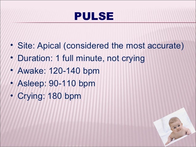
The apical pulse is a common arterial pulse site. It is the pulse measured over the chest where the heart’s mitral valve is best heard. In fact, it is the most efficient way to measure heart function. A stethoscope is used to measure the apical pulse, and it is best assessed while the patient is either lying down or sitting.
Apical pulse happens when the left ventricle of the heart contracts. Left ventricle forms the apex of the heart. Hence, the apical pulse is best measured by placing a stethoscope over the area of the apex. Doctors calculate the number of pulsations per minute. Measuring apical value is a noninvasive method.
What is Radial Pulse?
The radial pulse is one of the peripheral pulse sites. It is the pulse measured in the radial artery, which runs closer to the surface of the skin at the wrist. It gives the heartbeat per minute.
Radial pulse is assessed by placing three fingertips lightly upon the radial artery at the wrist. The exact location is one inch from the base of the thumb. In newborns and children under five, it is difficult to measure the radial pulse. Therefore, the apical pulse is usually measured in newborns and children.
What are the Similarities Between Apical and Radial Pulse?
- Radial and apical pulse are important to diagnose heart conditions.
- They are measured per minute and give the heart rate.
What is the Difference Between Apical and Radial Pulse?
The pulse measured at the apex of the heart is called the apical pulse, while the pulse at your wrist is called the radial pulse. So, this is the key difference between apical and radial pulse. The apical pulse is usually measured using a stethoscope while the radial pulse is measured using the tips of three central fingers.
Moreover, another difference between apical and radial pulse is that the apical pulse is the heart rate directly over the heart while the radial pulse gives the pulse on a location away from the heart. The radial pulse is difficult to measure in newborns and children below five while apical pulse can be measured in newborns and children below five.
Below infographic tabulates the differences between apical and radial pulse.
Summary – Apical vs Radial Pulse
Apical Pulse Area
Pulse or heart rate is a good indicator of heart health or the overall health of a person. The apical pulse is the pulse directly over the top of the heart. Measuring the apical value is noninvasive, and it is the best way to detect heart function. The radial pulse is one of the peripheral pulse sites in which the radial artery runs close to the skin of the inner wrist. In simple words, the radial pulse is the pulse at your wrist. Thus, this is the summary of the difference between apical and radial pulse.
Reference:
1. “How To Take Your Pulse”. Livescience.Com, 2020, Available here.
2. “Apical Pulse: Definition, Location, And More”. Healthline, 2020, Available here.
Image Courtesy:
1. “Doctor listening to patient’s heartbeat using stethoscope” (CC0) via Pikrepo
2. “Radial pulse” By U.S. Air Force Staff Sgt. Jeanette Copeland – Image # 040413-F-5646C-00 (description, full size link) (Domini públic) via Commons Wikimedia
Related posts:
Also found in: Dictionary, Thesaurus, Encyclopedia.
pulse
[puls] 2. the beat of the heart as felt through the walls of a peripheral artery, such as that felt in the radial artery at the wrist. Other sites for pulse measurement include the side of the neck (carotid artery), the antecubital fossa (brachial artery), the temple (temporal artery), the anterior side of the hip bone (femoral artery), the back of the knee (popliteal artery), and the instep (dorsalis pedis artery).What is felt is not the blood pulsing through the arteries (as is commonly supposed) but a shock wave that travels along the walls of the arteries as the heart contracts. This shock wave is generated by the pounding of the blood as it is ejected from the heart under pressure. It is analogous to the hammering sound heard in steam pipes as the steam is forced into the pipes under pressure. A pulse in the veins is too weak to be felt, although sometimes it is measured by sphygmograph (see below); the tracing obtained is called a phlebogram
 .
.The pulse is usually felt just inside the wrist below the thumb by placing two or three fingers lightly upon the radial artery. The examiner's thumb is never used to take a pulse because its own pulse is likely to be confused with that of the patient. Pressure should be light; if the artery is pressed too hard, the pulse will disappear entirely. The number of beats felt in exactly 1 minute is the pulse rate.
In taking a pulse, the rate, rhythm, and strength or amplitude of the pulse are noted. The average rate in an adult is between 60 and 100 beats per minute. The rhythm is checked for possible irregularities, which may be an indication of the general condition of the heart and the circulatory system.
The amplitude of a pulse can range from totally impalpable to bounding and full; however, such terms are vague and subject to misinterpretation. To provide a more standardized description of pulse amplitude some agencies and hospitals use a scale that provides a more objective evaluation and reporting of the force of a pulse. On such a scale zero would mean that the pulse cannot be felt; +1 would indicate a thready, weak pulse that is difficult to palpate, fades in and out, and is easily obliterated with slight pressure; +2 would be a pulse that requires light palpation but once located would be stronger than a +1; +3 would be considered normal; and a +4 pulse would be one that is strong, bounding, easily palpated, and perhaps hyperactive, and could indicate a pathological condition such as aortic regurgitation.
If a pulse is noted to be weaker during inhalation and stronger during exhalation (pulsus paradoxus), this could indicate either greater reduction in the flow of blood to the left ventricle than is normal, as in constrictive pericarditis or pericardial effusion, or a grossly exaggerated inspiratory maneuver, as in tracheal obstruction, asthma, or emphysema.
An instrument for registering the movements, form, and force of the arterial pulse is called a sphygmograph. The sphygmographic tracing (or pulse tracing) consists of a curve having a sudden rise (primary elevation) followed by a sudden fall, after which there is a gradual descent marked by a number of secondary elevations.
ap·i·cal pulse
(ap'i-kăl pŭlsApical Pulse Age
)apical pulse
Want to thank TFD for its existence? Tell a friend about us, add a link to this page, or visit the webmaster's page for free fun content.
Link to this page:
Apical Pulse Normal Range
